
Shop by Brand


The most distal bone in the horse’s leg is the coffin bone. This critical bone has other names, such as distal phalanx, third phalanx, or even P3 for the abbreviation fans. The coffin bone is the hoof shaped bone that attaches to the laminae in the hoof. The coffin joint is the intersection between the coffin bone and the next bone up, the short pastern bone. Which is also called the second phalanx or P2. Adding one more bone to the mix, the navicular bone sits behind the coffin bone and below the small pastern bone. There’s a patch of cartilage between the navicular bone and the coffin joint, and a patch of cartilage between the navicular bone and associated tendons. The navicular area also has a bursa, which is a sack of fluid that helps the tendons within the hoof glide around.
These structures and the coffin joint are within the hoof and support the weight of your horse. That’s a lotta weight for some small bones. Inside the hoof, the coffin bone attaches to the blood and nerve infused laminae layer. This sensitive layer connects to the insensitive laminae layer which connects to the hoof wall. Any trauma or disease to the hoof can severely affect the laminae and the bones inside the hoof.
There’s also the deep digital flexor tendon (DDFT) inside the hoof, which comes from the back of the leg, into the hoof, under the navicular bone, and attaches to the back of the coffin bone. The DDFT functions to flex the coffin joint, helping your horse walk.
There are so many structures in the hoof that many things can happen. Believe it or not - the coffin bone (or navicular bone for that matter) can fracture, despite the protection of the hoof. The coffin bone is also capable of becoming deformed due to the shape of the hoof. Think about a club foot or constricted heels that change the hoof shape. This can affect the coffin bone.
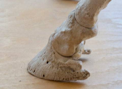
The navicular bone is just visible here, sitting right on top of the end of the coffin bone.

The coffin joint itself can become inflamed - much like a hock joint or a stifle joint. Wear and tear from repetitive motion creates inflammation. The navicular bursa can also become inflamed.
Osteitis, specifically pedal osteitis, is another issue to be aware of. Osteitis is a demineralization of the bone - and in the case of the coffin joint, is largely due to concussion, hard ground, frequent bruising. The coffin bone itself is damaged. Supportive farrier work, rest, and even medications are often warranted. This will cause lameness, and can be seen on X-rays.
And then you have the generalized navicular syndrome condition, often referring to a horse as “having navicular.” What causes this? Perhaps is the cartilage in the area degenerating, perhaps it’s a mechanical change, perhaps it’s an arthritic type of change. It’s been shown that navicular changes may also be related to toe first landing and other mechanical changes due to compression of the area and/or poor farrier care. Every horse will have a different set of circumstances.
The take away message from all of this is that your horse’s coffin bone and surrounding structures take the weight of your entire horse. Daily attention to the digital pulse, regular health check ups including lameness exams, regular farrier care, and proactive X-rays of the hoof can save your horse a lot of discomfort if issues are caught early.